Арсланова_Г_А_и_др_Essential_English_for_Biology_Students (1). Kazan federal university
 Скачать 7.01 Mb. Скачать 7.01 Mb.
|
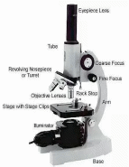
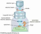
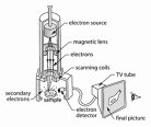
Text 2.3. Microscopes ■ Essential targets: By the end of this text you should be able to: ● describe the main features of a light microscope and an electron microscope ● distinguish between the terms magnification and resolving power ● give the approximate size of different biological structures using an appropriate unit of measurement. Pre-reading ■ Discuss these questions with your partner. Then find the answers in the text. 1. Who invented a microscope? 2. What types of microscopes are used today? ■ Read the given text and make your essential assignments: A  microscope is used to produce a magnified image of an object or specimen. Anton van Leeuwenhoek (1632-1723) was the first to invent a microscope powerful enough to explore the world of microbes. His discoveries stimulated an explosion of interest in the scientific use of microscopes. Since the 18th century many new types have been invented, of which the most commonly used today are the compound light microscope and the electron microscope. The compound light microscope The compound light microscope is also called a light microscope or optical microscope. The compound light microscope is also called a light microscope or optical microscope. The lenses refract (bend) the light to give a magnified image of the object. The image may be projected directly into the viewer’s eye or into photographic film. A photograph taken through a light microscope is called a photomicrograph or light micrograph. Magnification and resolution The magnification of an instrument is the increase in the apparent size of the object. The total magnification of a compound microscope is worked out by multiplying the magnification of the objective lens by that of the ocular lens. There is virtually no limit to the magnification produced by a light microscope; it depends on the power of the lenses used. However, above a certain magnification the image becomes blurred and it is impossible to distinguish structures lying close together. This limit of effective magnification is called the resolving power or resolution of the microscope. It is defined as the ability of a microscope to show two objects as separate. The resolving power of the light microscope is limited by the wavelength of light. Light microscopes can magnify objects up to about 1500 times without losing clarity. The electron microscope Electron microscopes use a beam of electrons instead of a beam of light. Electron beams have a much smaller wavelength than light rays, so electron microscopes have greater resolving powers and can produce much higher effective magnifications than light microscopes. There are two main types of electron microscopes: the transmission electron microscope (TEM), and the scanning electron microscope (SEM). The transmission electron microscope T  he TEM is used to study the details of the internal structure of cells. Extremely thin samples of the specimen are needed. To make these the specimen is supported in a resin block to prevent in collapsing during cutting, and is sliced with a diamond or glass knife. The section is then impregnated with a heavy-metal stain, such as osmium tetroxide. As the beam passes through the specimen, electrons are absorbed by heavily stained parts but pass readily through the lightly stained parts. Electromagnets bend the electron beam to focus an image onto a fluorescent screen or photographic film. Photograph taken through an electron microscope is called an electron micrograph. The most modern TEMs distinguish objects as small as 0.2nm. This means that they can produce clear images magnified up to 250 000 times. The magnification is varied by changing the strength of the electromagnets. The scanning electron microscope The SEM is used to produce three-dimensional images of the surface of specimens. Electron are reflected from the surface of a specimen stained with a heavy metal. This enables the SEM to produce images of whole specimens: cells, tissues, or even organisms. A  lthough electron microscopes have revolutionized cell biology, they have not completely replaced light microscopes. Light microscopes are used to examine living and unstained specimens. Preparation of specimens for electron microscopy is complicated and time-consuming. Electron microscopes are very expensive and can be used only to study dead specimens stained with heavy metal, which might well produce artifacts. ■ Glossary of essential terms for you to know.
■ Your Essential Assignments I. Quick check 1. How is the magnification varied in: a) a light microscope b) an electron microscope? 2. Why is the resolving power of an electron microscope so much better than of a light microscope? 3. What is the approximate size of the smallest structure that can be observed with a light microscope? II. Fill in the missing words:
III. Use monolingual English dictionary and write down what could the words given below mean: microscope, to refract, magnification, sample, ray. IV. Match these words with their definitions:
V. Find English equivalents to the following word combinations:
VI. Give Russian equivalents to the following English terms:
VII. Find synonyms among the pool of words:
VIII. Answer the following questions. Use all information given before: What are microscopes used for? What types of microscopes are most commonly used today? What is a compound light microscope? What does the magnification of an instrument depend on? How do electron microscopes differ from compound light microscopes? What are the main types of electron microscopes? What is the difference between the transmission electron microscope and the scanning electron microscope? IX. Match the sentence halves. Make complete centences:
X. Read and translate the short text without any dictionary: Fact of life: A new microscope, called a scanning tunneling microscope, was invented in 1980. It measures surface features by moving a sharp probe over the object’s surface. It can achieve magnifications of 100 million, allowing scientists to view atoms on the surface of a solid. This type of microscope is likely to have a major impact on biology. Recently, it has been used to view DNA directly. XI. Food for thought: Suggest which unit should be used when calculating the diameter of the DNA molecule. Why might there be a discrepancy between the actual diameter and that estimated from the scanning tunneling micrograph? ■ Have Some Fun! Biologist Jokes! A young college student stayed up all night studying for his zoology test the next day. As he entered the classroom, he saw ten stands with ten birds on them with a sack over each bird and only legs showing. He sat right on the front row because he wanted to do the best job possible. The professor announced that the test would be to look at each set of bird legs and give the common name, habitat, genus, species, etc. The student looked at each set of bird legs. They all looked the same to him. He began to get upset. He had stayed up all night studying, and now had to identify birds by their legs. The more he thought about it, the madder he got. Finally, he could stand it no longer. He went up to the professor`s desk and said: “What a stupid test! How could anyone tell the difference between birds by looking at their legs?” With that the student threw his test on the professor`s desk and walked out the door. The professor was surprised. The class was so big that he didn`t know every student`s name, so as the student reached the door the professor called: “Mister, what`s your name?” The enraged student pulled up his pant legs and said: “You guess, buddy! You guess!” |
