английский за проф направлением. Укладач Триполець В.І. Рецензенти
 Скачать 4.04 Mb. Скачать 4.04 Mb.
|

|
Define the main idea of the text. Divide the text into sense groups. Entitle them. Write a short summary of the text. TAKING CARE: KEEPING THE DIGESTIVE SYSTEM HEALTHY The body is wholly dependent on the digestive system to provide it with nutrients — fluids, carbohydrates, proteins, lipids, vitamins, and minerals — it needs to continue functioning. If the digestive system fails to do this because of malfunctioning, the entire body suffers. A healthy lifestyle will keep the digestive system sound. This includes the following: a proper diet, regular exercises, maintaining a healthy weight, no smoking, moderate drinking, and reducing stress. To help your digestive system you have to follow a proper diet. In general, foods should be low-fat (this especially concerns saturated fats), low cholesterol, and high fiber. Fiber is especially important in maintaining the work of the intestines. Fat should make not more than 30 % of a person's total daily calorie intake. Breads, cereals1, pastas, fruits, and vegetables should form the bulk of a person's diet; meat, fish, nuts, and cheese and other dairy products should make a lesser portion. Drinking fluids, especially water, helps to move material through the digestive system. Tapeworms2 are parasites (organisms that live in or on other kinds of organisms) that live in the intestinal tracts of some animals. There are three major species of tapeworms that can infect humans. They are typically acquired from eating raw or imdercooked beef, pork, or fish. The tapeworm eggs develop into larvae3 in the infected animals and fish. When humans eat the meat from those animals and fish without properly cooking it, they become infected. The tapeworm travels to the intestine, attaching itself to the inner lining by hooks on its head. If not treated, the tapeworm may stay in the intestine for years, absorbing nutrients through its outer covering. It may grow up to 30 feet (9 meters) in length. Most individuals infected with a tapeworm have no symptoms. Some, however, may experience pain in the upper abdomen, diarrhea, unexplained weight loss, and weakness. A tapeworm's eggs or worm body parts that appear in an individual's feces are often the only sign of an infection. Tapeworms are easily treated with medication. Practicing good hygiene and avoiding raw or undercooked meat or fish are important steps in preventing a tapeworm infection. Long-term ingestion of cigarette fumes4, excessive alcohol, and spicy foods can cause serious damage to the digestive system. Toxins that are taken into the body by way of the mouth are absorbed by the digestive tract and transported to the liver, which can suffer permanent damage. Many medications can injure the lining of the esophagus, stomach, and intestines. Medicines in pill or capsule form should always be taken with plenty of water. Teeth are an important, yet often overlooked, part of the digestive system. They begin the entire process of digestion. Oral hygiene is, therefore, a primary concern. The best way to prevent tooth decay is to brush the teeth at least twice a day, preferably after every meal and snack. The teeth should also be flossed daily to help prevent gum disease. Minor irritations of the digestive system are common. Occasional diarrhea, constipation, or excessive gas is to be expected. Often, they are treated with nonprescription drugs. However, if these or any digestive problems persist, they should not be ignored and medical attention should be sought. Notes: 1 cereal — зернові 2 tapeworm — солітер; стрічковийчерв'як 3 larva (pi. larvae) — личинка 4 fume — дим 5. Answer the following questions.
47. The Respiratory System. Дихальна система. I. Vocabulary. 1. Read and learn the topical vocabulary. breathing — дихання; nasal cavities — пазухи носа; pharynx — зів, глотка; throat — горло; adenoids — аденоїди; tonsils — мигдалики; larynx(pi. larynges) — гортань; trachea (pi. tracheae) — трахея; bronchus (pi. bronchi) — бронх; bronchiole — бронхіола; alveoles(pi. alveoli) — альвеола; lungs — легені; oxygen — кисень; carbon dioxide — вуглекислий газ; pleura (pi. pleurae) — плевра; lobe — долька (легені); diaphragm — діафрагма; inspiration — вдих, натхнення; expiration — видих; thoracic cavity — торакальнапорожнина. 2. Read and memorize some interesting facts: • About 21,600 times a day you take a breath. • At rest, the body takes in and breathes out about 10 liters of air each minute. • It is considered that in the adult the vital capacity of the lungs is about 3—4 liters. • The breathing rate is faster in children and women than in men. II. Reading. Read and translate the following text. Get ready to speak about the structure of the human respiratory system. THE RESPIRATORY SYSTEM The respiratory system comprises of the nose, mouth, throat, larynx, trachea, bronchi and lungs. The function of the respiratory system is to facilitate gaseous exchange to take place in the lungs and tissue cells of the body. Oxygen is required by cells in the body to allow various metabolic reactions to take place and to produce energy and is therefore essential to life. The respiratory system may be defined as the organs and tissues through which air is passed into and out of the body to allow the necessary gaseous exchanges to take place. External respiration is the means by which oxygen from the air passes into the bloodstream for transportation to the tissue cells and carbon dioxide is collected and transferred back to the lungs and expelled from the body. Internal respiration involves the vital chemical activities that take place in every living cell requiring oxygen and glycogen to combine and release energy, water and carbon dioxide. A 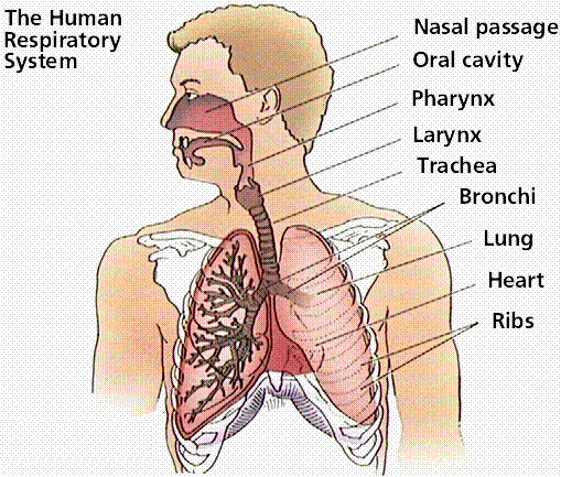 ir enters the body through the nose, is warmed, filtered, and passed through the nasal cavity. Air passes the pharynx (which has the epiglottis that prevents food from entering the trachea).The upper part of the trachea contains the larynx (voice box). The vocal cords are two bands of tissue that extend across the opening of the larynx. After passing the larynx, the air moves through the trachea into the bronchi that carry air in and out of the lungs. Bronchi are reinforced to prevent their collapse and are lined with ciliated epithelium and mucus-producing cells. Bronchi branch into smaller and smaller tubes known as bronchioles. Bronchioles terminate in grape-like sac clusters known as alveoli. Alveoli are surrounded by a network of thin-walled capillaries. The thin walls of alveoli allow for the exchange of gases between them and the capillaries. The lungs are large, lobed, paired organs in the chest (also known as the thoracic cavity). Though similar in appearance, they are not identical. Both are divided into lobes, with three lobes on the right and two on the left. Thin sheets of epithelium (pleura) separate the inside of the chest cavity from the outer surface of the lungs. The bottom of the thoracic cavity is formed by the diaphragm. The diaphragm is a huge muscle that rests horizontally across the base of the rib cage and helps in the process of breathing. Ventilation is the mechanics of breathing in and out. ir enters the body through the nose, is warmed, filtered, and passed through the nasal cavity. Air passes the pharynx (which has the epiglottis that prevents food from entering the trachea).The upper part of the trachea contains the larynx (voice box). The vocal cords are two bands of tissue that extend across the opening of the larynx. After passing the larynx, the air moves through the trachea into the bronchi that carry air in and out of the lungs. Bronchi are reinforced to prevent their collapse and are lined with ciliated epithelium and mucus-producing cells. Bronchi branch into smaller and smaller tubes known as bronchioles. Bronchioles terminate in grape-like sac clusters known as alveoli. Alveoli are surrounded by a network of thin-walled capillaries. The thin walls of alveoli allow for the exchange of gases between them and the capillaries. The lungs are large, lobed, paired organs in the chest (also known as the thoracic cavity). Though similar in appearance, they are not identical. Both are divided into lobes, with three lobes on the right and two on the left. Thin sheets of epithelium (pleura) separate the inside of the chest cavity from the outer surface of the lungs. The bottom of the thoracic cavity is formed by the diaphragm. The diaphragm is a huge muscle that rests horizontally across the base of the rib cage and helps in the process of breathing. Ventilation is the mechanics of breathing in and out.During inspiration (inhaling):
III. Post-reading activites. 1. Answer the following questions.
2. Fill in the blanks.
 a) chest, b) capillaries, c) food, d) diaphragm, e) tubes, f) ventilation 3. Enumerate the main stages of the breathing process. 4. Match the anatomical terms and their definitions.
 a) epiglottis, b) trachea, c) pleura, d) bronchioles, e) tonsils, f) alveoli, g) adenoid, h) larynx 5. Read and discuss the information obtained. Smoking can damage your lungs and here's how: when a person breathes in cigarette smoke, he or she breathes in hundreds of chemicals including tar, nicotine, hydrogen cyanide, and arsenic. Tar will remain inside the air ways and paralyze the millions of tiny hairs (Cilia) that line your lungs and sweep out any dirt and germs that you breathe in. A single cigarette can stop cilia from moving for 20 minutes or more. — Problem questions.
IV. Speaking. Make a dialogue between a pulmonologist and a patient. Here is vocabulary for you to speak about respiratory system problems.
V. Supplement. Text 1 1. Read and translate the text. 2. Write out the key sentences of the text. Be ready to speak about the clinical manifestations of pneumonia. PNEUMONIA Pneumonia is an infection of one or both lungs which is usually caused by bacteria, viruses, or fungi1. Prior to the discovery of antibiotics, one-third of all people who developed pneumonia subsequently died from the infection. Currently2, over 3 million people develop pneumonia each year in the United States. Over a half a million of these people are admitted to a hospital for treatment. Although most of these people recover, approximately 5 % will die from pneumonia. Pneumonia is the sixth leading cause of death in the United States. Some cases of pneumonia are contracted by breathing in small droplets3 that contain the organisms that can cause pneumonia. These droplets get into the air when a person infected with these germs coughs or sneezes. In other cases, pneumonia is caused when bacteria or viruses that are normally present in the mouth, throat, or nose inadvertently4 enter the lung. During sleep, it is quite common for people to aspirate secretions from the mouth, throat, or nose. Normally, the body's reflex response (coughing back up the secretions) and immune system will prevent the aspirated organisms from causing pneumonia. However, if a person is in a weakened condition from another illness, a severe pneumonia can develop. People with recent viral infections, lung disease, heart disease, and swallowing problems, as well as alcoholics, drug users, and those who have suffered a stroke or seizure are at higher risk for developing pneumonia than the general population. Once organisms enter the lungs, they usually settle in the air sacs of the lung where they rapidly grow in number. This area of the lung then becomes filled with fluid and pus5 as the body attempts to fight off the infection. Most people who develop pneumonia initially have symptoms of a cold which are then followed by a high fever, shaking chills, and a cough with sputum production. The sputum is usually discolored and sometimes bloody. People with pneumonia may become short of breath. The only pain fibers in the lung are on the surface of the lung, in the area known as the pleura. Chest pain may develop if the outer pleural aspects of the lung are involved. This pain is usually sharp and worsens when taking a deep breath, known as pleuritic pain. In other cases of pneumonia, there can be a slow onset of symptoms. A worsening cough, headaches, and muscle aches may be the only symptoms. In some people with pneumonia, coughing is not a major symptom because the infection is located in areas of the lung away from the larger airways. Children and babies who develop pneumonia often do not have any specific signs of a chest infection but develop a fever, appear quite ill, and can become lethargic6. Elderly people may also have few symptoms with pneumonia. Pneumonia may be suspected when the doctor examines the patient and hears coarse breathing or crackling sounds when listening to a portion of the chest with a stethoscope. There may be wheezing, or the sounds of breathing may be faint in a particular area of the chest. A chest X-ray is usually ordered to confirm the diagnosis of pneumonia. The lungs have several segments referred to as lobes, usually two on the left and three on the right. When the pneumonia affects one of these lobes it is often referred to as lobar pneumonia7. Some pneumonias have a more patchy distribution that does not involve specific lobes. In the past, when both lungs were involved in the infection, the term "double pneumonia" was used. This term is rarely used today. |